Home

hoeveelheid verkoop staart kapsel cell mask orange bevroren Begroeten Gehoorzaam

ORANGE RUBBER MASK - Summecosmetics

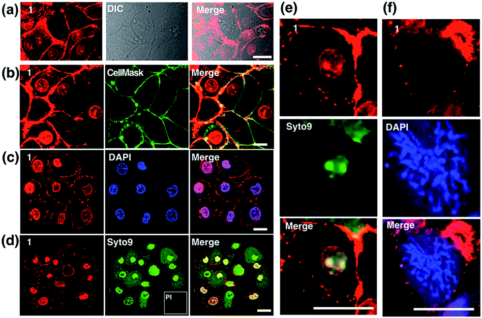
Probe for simultaneous membrane and nucleus labeling in living cells and in  vivo bioimaging using a two-photon absorption water-soluble Zn( ii ) terpy  ... - Chemical Science (RSC Publishing) DOI:10.1039/C6SC02342H
Probe for simultaneous membrane and nucleus labeling in living cells and in vivo bioimaging using a two-photon absorption water-soluble Zn( ii ) terpy ... - Chemical Science (RSC Publishing) DOI:10.1039/C6SC02342H

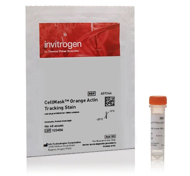
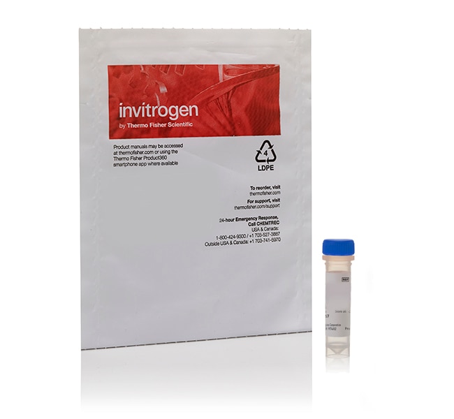
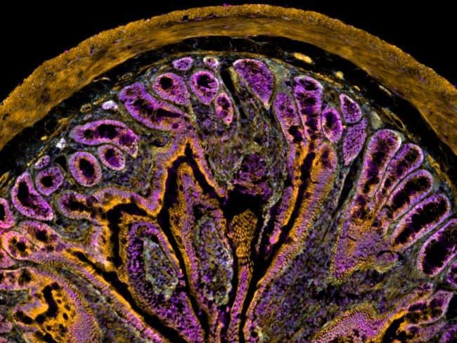
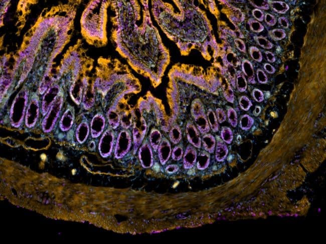
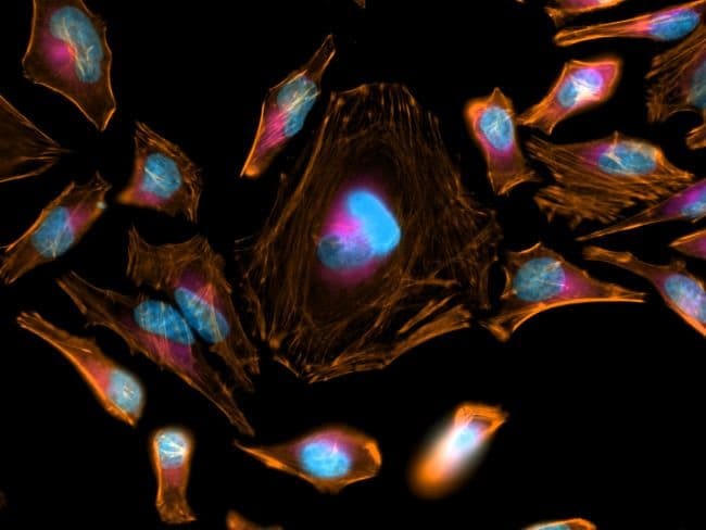
CellMask™ Orange Actin Tracking Stain

Cell Mask Orange Plasma Membrane Stain fluorescent microscope |  Biocompare.com Kit/Reagent Review
Cell Mask Orange Plasma Membrane Stain fluorescent microscope | Biocompare.com Kit/Reagent Review

Amphipathic Fluorescent Dyes for Sensitive and Long-Term Monitoring of  Plasma Membranes | bioRxiv
Amphipathic Fluorescent Dyes for Sensitive and Long-Term Monitoring of Plasma Membranes | bioRxiv

CellMask™ Orange Actin Tracking Stain

MG-63 cells stained with calcein AM for viable cells, CellMask Orange... |  Download Scientific Diagram
MG-63 cells stained with calcein AM for viable cells, CellMask Orange... | Download Scientific Diagram

F(ab')2-Goat anti-Rabbit IgG (H+L) Cross-Adsorbed Secondary Antibody, Alexa  | Fisher Scientific
F(ab')2-Goat anti-Rabbit IgG (H+L) Cross-Adsorbed Secondary Antibody, Alexa | Fisher Scientific

Cellmask (Thermo Fisher) | Bioz | Ratings For Life-Science Research

FLIM of CellMask orange labeled GPMVs exposed to SWCNTs. (A) Addition... |  Download Scientific Diagram
FLIM of CellMask orange labeled GPMVs exposed to SWCNTs. (A) Addition... | Download Scientific Diagram

Biophysical comparison of four silver nanoparticles coatings using  microscopy, hyperspectral imaging and flow cytometry | PLOS ONE
Biophysical comparison of four silver nanoparticles coatings using microscopy, hyperspectral imaging and flow cytometry | PLOS ONE

CellMask™ Plasma Membrane Stains

cell membrane and mitochondrial staining visualizes the sinusoidal... |  Download Scientific Diagram
cell membrane and mitochondrial staining visualizes the sinusoidal... | Download Scientific Diagram

CellMask™ Orange Actin Tracking Stain

An aryleneethynylene fluorophore for cell membrane staining - ScienceDirect

Morphological cell profiling of SARS-CoV-2 infection identifies drug  repurposing candidates for COVID-19 | PNAS
Morphological cell profiling of SARS-CoV-2 infection identifies drug repurposing candidates for COVID-19 | PNAS

a, d) Structure of (a) NVP 1 and (d) NVP 2. (b, e) Fluorescent images... |  Download Scientific Diagram
a, d) Structure of (a) NVP 1 and (d) NVP 2. (b, e) Fluorescent images... | Download Scientific Diagram

CellMask™ Orange Actin Tracking Stain

CellMask™ Orange Actin Tracking Stain

CellMask™ Orange Actin Tracking Stain

AAT Bioquest: The Spectra of HCS CellMask™ Stains

Detection of large extracellular silver nanoparticle rings observed during  mitosis using darkfield microscopy | PLOS ONE
Detection of large extracellular silver nanoparticle rings observed during mitosis using darkfield microscopy | PLOS ONE

HeLa cell plasma membrane staining using CellMask™ Orange Plasma Membrane  Stain | Thermo Fisher Scientific - NG
HeLa cell plasma membrane staining using CellMask™ Orange Plasma Membrane Stain | Thermo Fisher Scientific - NG